

A Dna Nucleotide May Contain
Chapter 5. Nucleotides & Nucleic Acids
- 5.1 Nucleotides and the Phosphodiester Bail
- 5.2 Dna: DNA
- five.3 Ribonucleic acid: RNA
Introduction
Nucleic acids are macromolecules made upward of monomers called nucleotides. They are the near of import macromolecules for the continuity of life. They bear the genetic data of a cell and instructions for the functioning of the cell. Nucleic acids are information molecules that serve as blueprints for the proteins that are made past cells. They are also the hereditary cloth in cells, as reproducing cells pass the blueprints on to their offspring.
The two main types of nucleic acids are deoxyribonucleic acid (DNA) and ribonucleic acid (RNA). Deoxyribonucleic acid is the genetic material found in all living organisms. It is found in the nucleus of eukaryotes and in the chloroplasts and mitochondria. In prokaryotes, the DNA is not enclosed in a nucleus.
The unabridged genetic content of a jail cell is known as its genome. In eukaryotic cells, DNA forms a complex with histone proteins to form chromatin, the substance of eukaryotic chromosomes. A chromosome may incorporate tens of thousands of genes. Many genes contain the information to brand protein products; other genes code for RNA products. Dna controls all of the cellular activities past turning the genes "on" or "off."
The other type of nucleic acid, RNA, is mostly involved in poly peptide synthesis. DNA molecules utilize an intermediary, called messenger RNA (mRNA), to communicate with the rest of the cell. Other types of RNA, such as rRNA, tRNA, and microRNA, are involved in protein synthesis and its regulation.
5.1 | Nucleotides and the Phosphodiester Bond
By the end of this section, you lot will be able to:
- Identify the three components of nucleotide structure.
- Recognize how nucleotides and nucleic acids are related.
- Proper noun the type of bond that holds nucleotides together & identify it in a nucleic acid structure.
Dna and RNA are fabricated up of monomers known as nucleotides. The nucleotides combine with each other to form a nucleic acrid, DNA or RNA. Each nucleotide is made upwardly of three components: a nitrogenous base of operations, a pentose (five-carbon) sugar, and a phosphate group (Figure five.ii). Each nitrogenous base in a nucleotide is fastened to a sugar molecule, which is fastened to one or more than phosphate groups.
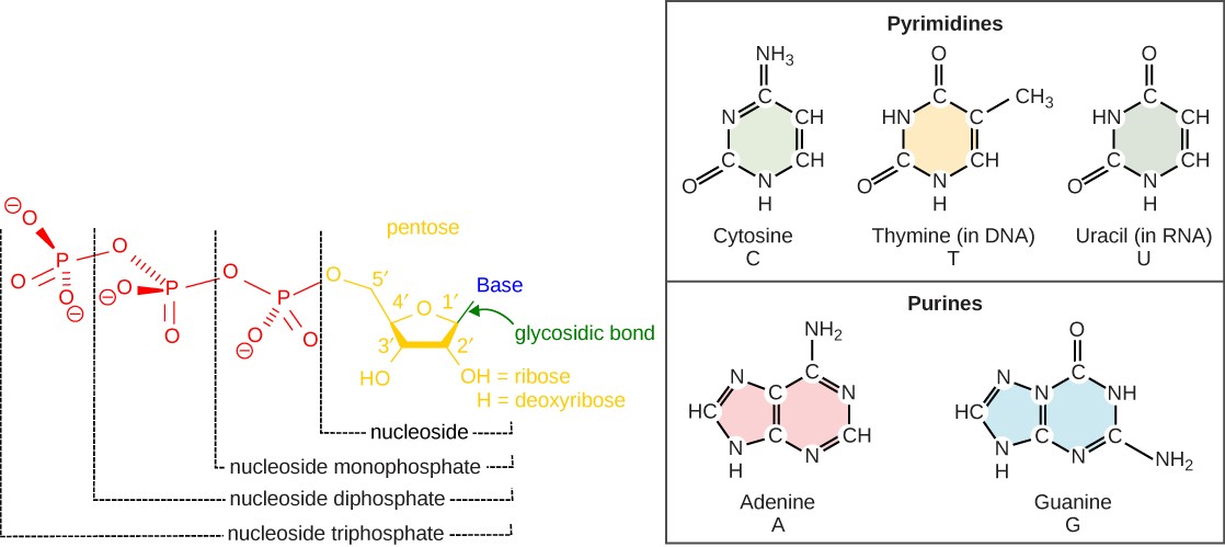
The nitrogenous bases are organic molecules that contain nitrogen. They are bases because they contain an amino group that has the potential of bounden an extra hydrogen. Each nucleotide in Dna contains one of iv possible nitrogenous bases: adenine (A), guanine (G) cytosine (C), and thymine (T). Each nucleotide in RNA contains one of four possible nitrogenous bases: adenine (A), guanine (Chiliad) cytosine (C), and uracil (U). Adenine and guanine are classified every bit purines and have ii carbon-nitrogen rings. Cytosine, thymine, and uracil are classified as pyrimidines, which take a single carbon-nitrogen ring (Figure 5.2).
The carbon atoms of the pentose sugar molecule in each nucleotide are numbered as 1′, 2′, 3′, four′, and 5′ (1′ is read as "one prime"). The nitrogenous base is attached to the 1′ carbon and the phosphate group is fastened to the hydroxyl group of the 5′ carbon. In RNA, the pentose sugar is ribose, which has a hydroxyl group fastened to the 2′ carbon. In DNA, the pentose sugar is deoxyribose, which has a hydrogen atoms attached to the 2′ carbon. The "deoxy" in the name of Deoxyribonucleic acid refers to the missing oxygen atom at the 2′ carbon (Figure 5.2).
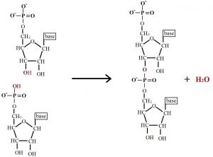
Nucleic acids are long, linear chains of nucleotides. Phosphodiester linkages are covalent bonds betwixt the iii′ carbon of i nucleotide and the 5′ phosphate group of some other. They form past dehydration synthesis reactions (Effigy v.3). Nucleic acids have directionality: the outset nucleotide in the chain has a free phosphate group at the 5′ end of the molecule. The last nucleotide added has a free 3′ hydroxy group at the 3′ cease of the molecule. Nucleotides are always added on to the 3′ finish.
5.2 | Deoxyribonucleic Acid: Deoxyribonucleic acid
By the end of this section, you will exist able to:
- Describe the structure and role of DNA.
- Talk over the similarities and differences between eukaryotic and prokaryotic Dna.
v.2.ane The Double Helix
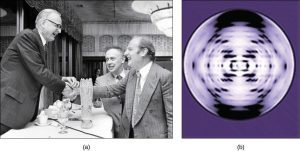
In the 1950s, Francis Crick and James Watson worked together to determine the structure of DNA at the Academy of Cambridge, England. Other scientists like Linus Pauling and Maurice Wilkins were too actively exploring this field. Pauling had discovered the secondary structure of proteins using Ten-ray crystallography. In Wilkins' lab, researcher Rosalind Franklin was using Ten-ray diffraction methods to understand the structure of Deoxyribonucleic acid. Watson and Crick were able to piece together the puzzle of the DNA molecule on the footing of Franklin's data because Crick had also studied X- ray diffraction (Figure 5.iv). In 1962, James Watson, Francis Crick, and Maurice Wilkins were awarded the Nobel Prize in Medicine. Unfortunately, by then Franklin had died, and Nobel prizes are not awarded posthumously.
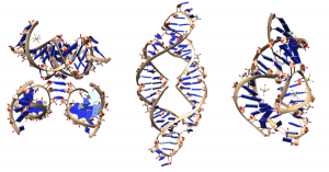
Watson and Crick correctly proposed that Dna is made up of two strands that are twisted around each other to form a correct-handed helix. Ii strands of nucleotides are held together by hydrogen bonds that form betwixt pairs of nitrogenous bases. The sugar and phosphate "backbone" forms the outside of the helix. The nitrogenous bases are stacked in the interior, like the steps of a ladder. The two strands are anti-parallel in nature; that is, the three′ stop of ane strand faces the five′ end of the other strand (Figure five.5).
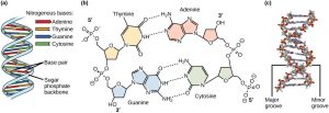
Only certain types of base pairing occur. A can only pair with T, and Yard can only pair with C, as shown in Figure five.v. This is known as the base of operations complementary rule. In other words, the DNA strands are complementary to each other. If the sequence of one strand is 5′-AATTGGCC-iii′, the complementary strand would accept the sequence iii′-TTAACCGG-v′. The fact that the 2 strands of a DNA molecule are complementary allows DNA to replicate. During DNA replication, each strand is copied, resulting in a daughter DNA double helix containing one parental DNA strand and a newly synthesized strand. The base pairs are stabilized by hydrogen bonds; adenine and thymine form ii hydrogen bonds and cytosine and guanine form iii hydrogen bonds.
A mutation occurs, and cytosine is replaced with adenine. What affect do you think this will take on the Deoxyribonucleic acid structure?
5.2.2. DNA Packaging in Cells
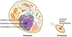
When comparing prokaryotic cells to eukaryotic cells, prokaryotes are much simpler than eukaryotes in many of their features (Figure v.half dozen). Most prokaryotes incorporate a single, circular chromosome that is found in an expanse of the cytoplasm chosen the nucleoid.
The size of the genome in one of the most well-studied prokaryotes, Due east. coli, is 4.6 meg base of operations pairs (approximately 1.1 mm, if cut and stretched out). And so how does this fit inside a small bacterial prison cell? The Dna is twisted by what is known every bit supercoiling. Supercoiling ways that Dna is either under-wound (less than one turn of the helix per 10 base pairs) or over-wound (more than than 1 plough per 10 base of operations pairs) from its normal relaxed state. Some proteins are known to be involved in the supercoiling; other proteins and enzymes, such as Deoxyribonucleic acid gyrase, help in maintaining the supercoiled structure.
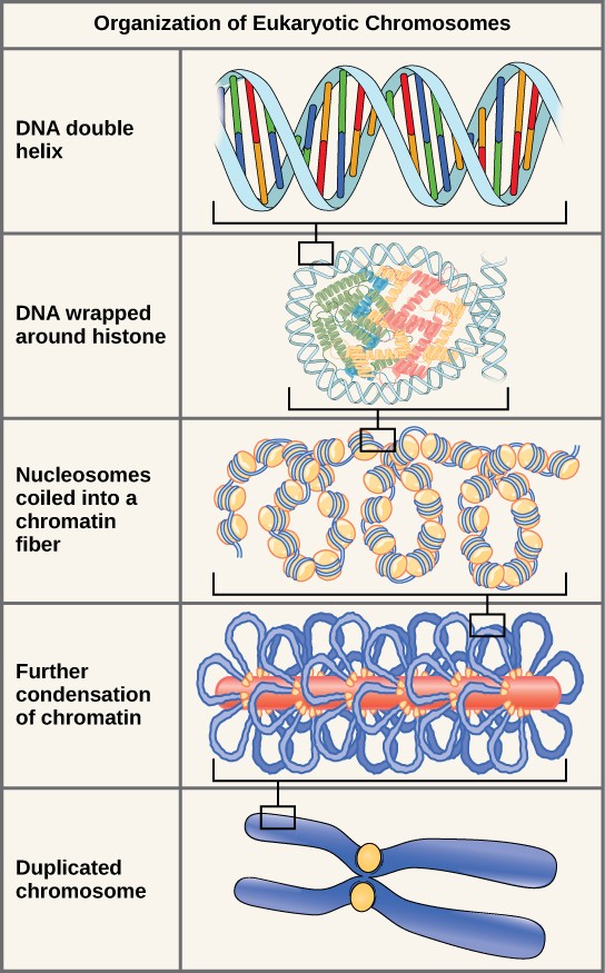
Eukaryotes, whose chromosomes each consist of a linear Dna molecule, employ a different type of packing strategy to fit their DNA within the nucleus (Figure 5.vii). At the virtually basic level, DNA is wrapped around proteins known every bit histones to course structures chosen nucleosomes. The histones are evolutionarily conserved proteins that are rich in basic amino acids and form an octamer. The DNA (which is negatively charged because of the phosphate groups) is wrapped tightly around the histone core. This nucleosome is linked to the next one with the assist of a linker DNA. This is too known every bit the "chaplet on a string" structure. This is farther compacted into a 30 nm fiber, which is the diameter of the structure. At the metaphase stage, the chromosomes are at their most compact, are approximately 700 nm in width, and are found in association with scaffold proteins.
In interphase, eukaryotic chromosomes have 2 distinct regions that can exist distinguished by staining. The tightly packaged region is known as heterochromatin, and the less dense region is known equally euchromatin. Heterochromatin normally contains genes that are not expressed (not actively transcribed to make a product), and is found in the regions of the centromere and telomeres. The euchromatin usually contains genes that are transcribed, with DNA packaged around nucleosomes but not further compacted.
5.3 | Ribonucleic Acid: RNA
Past the finish of this section, you volition be able to:
- Explain the structure and roles of RNA.
- Compare and dissimilarity the 2 types of nucleic acids.
Ribonucleic acid, or RNA, is mainly involved in protein synthesis. Like Deoxyribonucleic acid, RNA is made of nucleotides linked past phosphodiester bonds. Even so, the nucleotides in RNA incorporate ribose sugar instead of deoxyribose and the nitrogenous base uracil (U) instead of thymine (T). Unlike Dna, RNA is usually single-stranded. Still, most RNAs testify internal base pairing betwixt complementary sequences, creating a three-dimensional structure essential for their role.
There are four major types of RNA: messenger RNA (mRNA), ribosomal RNA (rRNA), transfer RNA (tRNA), and microRNA (miRNA). mRNA carries a copy of the genetic code from Deoxyribonucleic acid. If a cell requires a certain protein to be synthesized, the gene is turned "on" and the respective messenger RNA is synthesized. The RNA sequence is complementary to the sequence of the Deoxyribonucleic acid (except U replaces T). If the DNA strand has a sequence 5′-AATTGCGC-three′, the sequence of the complementary RNA is 3′-UUAACGCG-5′. The mRNA so interacts with ribosomes and other cellular mechanism and then that a protein can be fabricated from the coded message. The mRNA is read in sets of iii bases known equally codons. Each codon codes for a single amino acid.
Thus, information flow in an organism goes from DNA to mRNA to protein. DNA dictates the sequence of mRNA in a process known as transcription, and RNA dictates the structure of protein in a procedure known equally translation. This is known equally the Key Dogma of Molecular Biological science.
rRNA is a major constituent of ribosomes, to which the mRNA binds to make a protein product. tRNA carries the right amino acrid to the site of protein synthesis. miRNAs play a role in the regulation of gene expression. Tabular array 4.ii summarizes features of Deoxyribonucleic acid and RNA.
Tabular array 4.2 Features of DNA and RNA
| Dna | RNA | |
| Function | Carries genetic information | Involved in protein synthesis and regulation of cistron expression |
| Location | Remains in the nucleus | Leaves the nucleus |
| Structure | Double helix | Usually single-stranded |
| Sugar | Deoxyribose | Ribose |
| Pyrimidines | Cytosine, thymine | Cytosine, uracil |
| Purines | Adenine, guanine | Adenine, guanine |

A Dna Nucleotide May Contain,
Source: https://rwu.pressbooks.pub/bio103/chapter/nucleotides-and-nucleic-acids/
Posted by: ortegaandutimmose.blogspot.com
0 Response to "A Dna Nucleotide May Contain"
Post a Comment